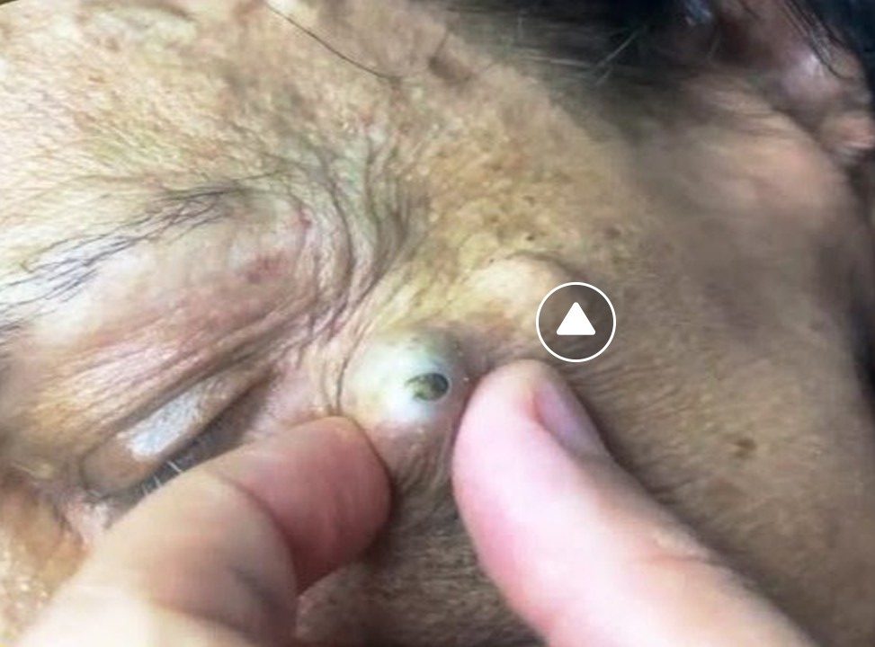
Horn-like skin conditions, often referred to as cutaneous horns (or cornu cutaneum), are unusual growths that resemble animal horns due to their conical, hard, and keratinous structure. These growths are composed of compacted keratin, the protein found in nails, hair, and the outer layer of skin. While they may appear alarming, cutaneous horns can arise from a range of underlying conditions, from benign to malignant. Below is a comprehensive explanation of these growths, their causes, symptoms, diagnosis, and treatment, based on current understanding and available evidence.
What Are Cutaneous Horns?
A cutaneous horn is a clinical term for a hard, conical projection from the skin that resembles an animal horn, though it lacks a bony core. These growths are typically:
- Appearance: Funnel-shaped, curved, or spike-like, with a yellowish-brown, whitish, or skin-colored hue. They may range from a few millimeters to several centimeters in length, with rare cases reaching up to 25 cm (e.g., the historical case of Madame Dimanche in the 19th century).
- Texture: Extremely hard due to hyperkeratosis (excessive keratin production).
- Location: Most commonly found on sun-exposed areas like the face, ears, nose, forearms, and hands, though they can appear anywhere, including less common sites like the penis or palmar aspect of fingers.
- Prevalence: More frequent in older adults (ages 60–80), with a slight predominance in men for malignant cases. They are less common in darker skin tones but can occur across all skin types.

Causes and Underlying Conditions
Cutaneous horns arise from excessive keratin growth (hyperkeratosis) triggered by various skin lesions. The underlying cause determines whether the horn is benign, precancerous, or cancerous. Approximately 60% of cutaneous horns are benign, while 20–40% are associated with premalignant or malignant conditions.
1. Benign Causes
- Seborrheic Keratosis: A common, noncancerous growth in older adults, often waxy or wart-like. It’s the most frequent benign cause of cutaneous horns.
- Viral Warts: Caused by human papillomavirus (HPV, especially HPV-2 subtype), these can lead to horn-like growths, particularly in children.
- Dermatofibroma: A firm, benign nodule, typically on arms or legs, that may develop a horn due to keratin buildup.
- Other: Molluscum contagiosum, nevi (moles), or psoriasis can rarely cause horns.
2. Precancerous Causes
- Actinic Keratosis: A scaly, sun-induced lesion that is the most common precancerous cause. About 5–10% of actinic keratoses may progress to squamous cell carcinoma (SCC).
- Bowen’s Disease: A form of squamous cell carcinoma in situ (non-invasive).
3. Cancerous Causes
- Squamous Cell Carcinoma (SCC): The most common malignant cause, especially in sun-exposed areas. SCC is found in up to 37% of penile cutaneous horns.
- Basal Cell Carcinoma (BCC): Less common but possible, often appearing as a pearly bump.
- Malignant Melanoma: Rare but serious, requiring immediate attention.
Risk Factors
- Sun Exposure: Chronic UV radiation is a primary trigger, explaining why horns often appear on the face, hands, and forearms.
- Age: Higher incidence in those over 60 due to cumulative sun damage and cellular aging.
- Skin Type: More common in fair-skinned individuals (phototypes I and II).
- Other: Trauma, burn scars, or HPV infection may contribute. Some cases in Asian populations suggest a link to warm climates.
Symptoms
Cutaneous horns are often asymptomatic, but symptoms may include:
- A hard, horn-like growth that may be curved or straight.
- Pain or inflammation if injured or infected.
- Rapid growth, discoloration (red, pink, or purple), or a wide base, which may indicate malignancy.
- Surrounding skin may be normal, thickened, or erythematous (red), with erythema suggesting cancerous potential.
Rarely, giant horns can impair function (e.g., hand movement) or become a cosmetic concern.
Diagnosis
Diagnosis involves:
- Clinical Examination: A doctor assesses the horn’s appearance, size, and location. Terrace-like ridges on the horn’s side suggest benign causes, while base erythema or rapid growth raises malignancy concerns.
- Biopsy: The gold standard. The entire horn is typically removed and examined histologically to identify the underlying lesion and rule out cancer. A deep partial biopsy may suffice for smaller lesions.
- Histopathology: Reveals hyperkeratosis with compact keratin. Benign lesions show orderly keratin layers, while malignant ones exhibit erratic growth.
Cutaneous horns may be confused with ectopic nails, necessitating histopathological confirmation.
Treatment
Treatment focuses on removing the horn and addressing the underlying lesion. Options include:
- Surgical Excision: The most common method, especially for suspected malignancy. The horn and base are removed with appropriate margins, and the defect may be closed primarily or with skin grafting for larger areas.
- Cryotherapy: Liquid nitrogen freezes precancerous lesions like actinic keratosis.
- Curettage and Electrodesiccation: Scraping and burning the lesion, often for benign or precancerous growths.
- Topical Treatments: For precancerous lesions, 5-fluorouracil or imiquimod may be used.
- Laser Therapy: Carbon dioxide or Nd:YAG lasers for precise removal, though less common.
If cancer is detected, further treatment (e.g., wider excision, radiation, or lymph node evaluation) depends on the type, size, and spread.
- Recovery: Varies by lesion size and type. Scarring is common, and horns may recur if the underlying cause persists.
- Prognosis: Excellent for benign lesions; good for early-detected cancers like SCC, but worse if metastasis occurs.
Prevention and Management
While no definitive prevention exists, you can reduce risk by:
- Sun Protection: Use high-SPF sunscreen, wear protective clothing, and avoid peak UV hours.
- Hydration and Stress Management: Adequate water intake (8 glasses daily) and stress-reducing practices like yoga may support overall skin health.
- Avoid Smoking: Tobacco impairs skin healing and may worsen lesions.
- Regular Skin Checks: Monthly self-exams and annual dermatologist visits help detect changes early.
Notable Cases
- Historical: In 1588, Margeret Gryffith, an elderly Welsh woman, was documented with a cutaneous horn. In the 19th century, Madame Dimanche grew a 25-cm horn from her forehead.
- Modern: In 2010, Zhang Ruifang (China, age 101) developed a 6-cm horn on her forehead, dubbed “Devil’s Horns.” In 2015, Liang Xiuzhen (China, age 87) grew a 13-cm horn, nicknamed “Unicorn Woman.”
When to Seek Medical Attention
Consult a healthcare professional if you notice:
- A new or changing horn-like growth.
- Pain, rapid growth, discoloration, or inflammation.
- A wide base or surrounding erythema.
Early evaluation is critical due to the risk of underlying malignancy.
Conclusion
Cutaneous horns are striking but uncommon skin growths caused by excessive keratin buildup from benign, precancerous, or cancerous lesions. While often harmless, their association with skin cancer (especially squamous cell carcinoma) necessitates prompt medical evaluation. Sun exposure is a key risk factor, making prevention through UV protection essential. With timely diagnosis and treatment, outcomes are generally favorable, but vigilance is crucial to address potential malignancies.


